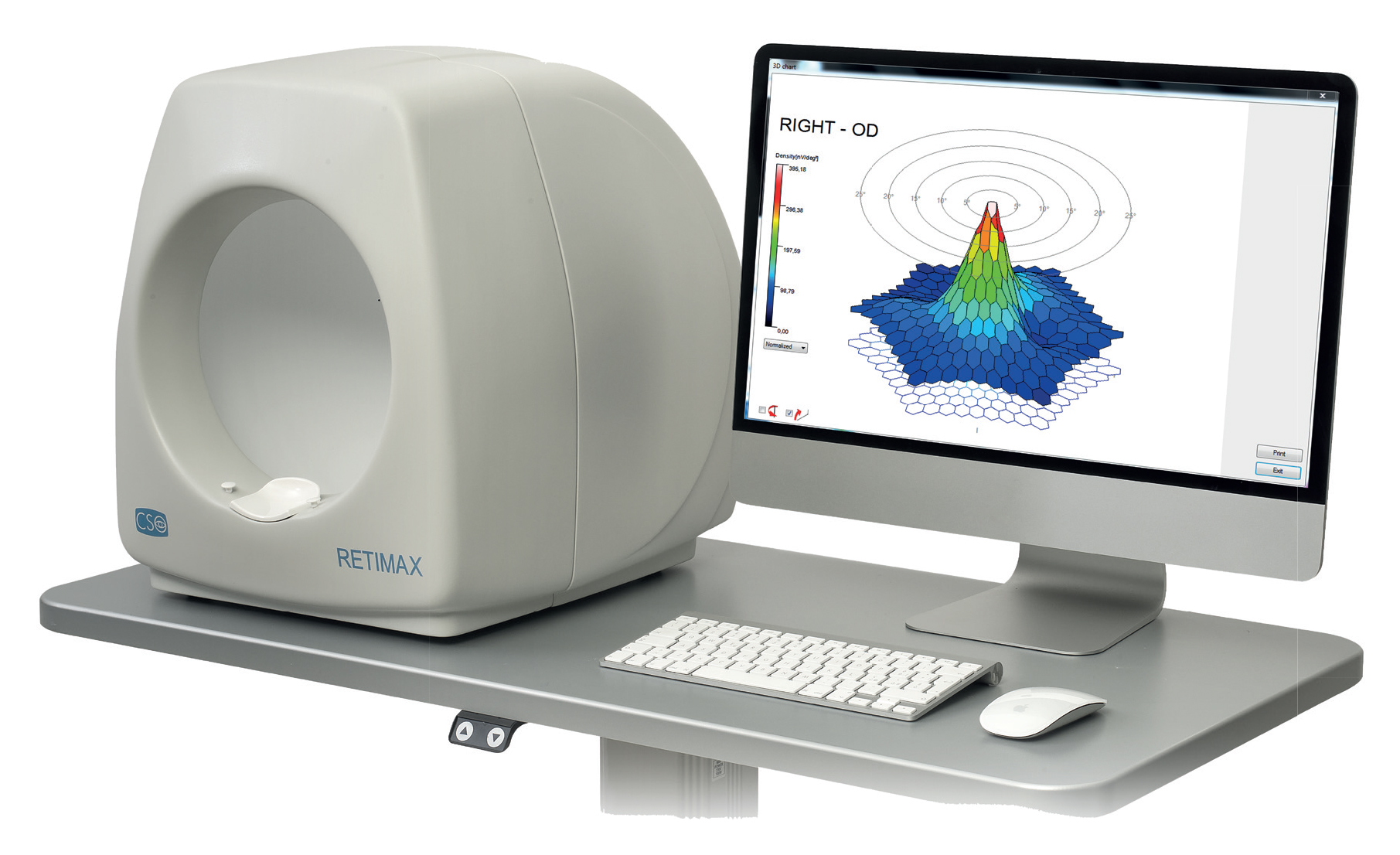
The following can be mapped:
-
Glaucoma
-
Age Related Macular Degeneration (ARMD)
-
Pigmented Retinitis
-
Scotomas of few millimeters in diameter
The extension of retinal dysfunction is quantified very accurately, particularly in the early stages of disease.
Retimax Advanced Plus, provides the newest total length binary M-sequence in real-time (Patent Pending) up to 82°. Visual angle, from 7 to 241 stimuli of retinal fields, in order to detect the objective bioelectrical response of each stimulated retinal field. Thanks to the innovative features of Retimax Advanced Plus, it is possible to display, in real time, the results of each stimulated retinal field. It provides the ophthalmologist the possibility of interacting directly with the patient during the test for best co-operation. This avoids artifacts or attention loss during the test. Retimax Advanced Plus provides age correlated normative data for multifocal ERG, PERG and VEP In order to compare the patient examined with the normal control group.
A number of stimulus settings are available for the user as:
-
Automated adjustment of subtended visual angle depending on the optical correction applied to the patient
-
Number of stimulus fields
-
Distortion of stimulated areas
-
Eccentricity
-
Checkerboards
-
Colours
The advanced analysis strategy includes:
-
Traces array
-
3D array
-
2D array
-
Retinal rings analysis
-
Quadrants analysis
-
Hemifield analysis (Patent Pending)
-
User defined personal settings
Additionally, Retimax Advanced Plus provides connections with slit lamps, fundus cameras and laser scanning ophthalmoscope OCT, in order to obtain, simultaneously, the detection of functional test and retinal image, or RNFL (Retinal Nerve Fiber Layer). Results of analysis strategy are printed out on a high-resolution colour printer. Files of graphics and text data can be exported to other programs for statistic analysis. A large range of electrodes are available as HKLOOP ring, fiber or contact lens for the patients best comfort during the examination. Retimax Advanced Plus has been designed to meet the international standard ISCEV (International society for Clinical Electrophysiology of Vision).
Retimax Advanced Plus configuration includes Ganzfeld flash, pattern stimulator with chin rest (up to 82°), electrodes set, personal computer, windows operating system or higher, USB port connection, ink jet printer, table.


