Electrophysiology
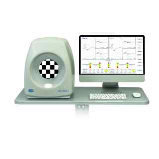
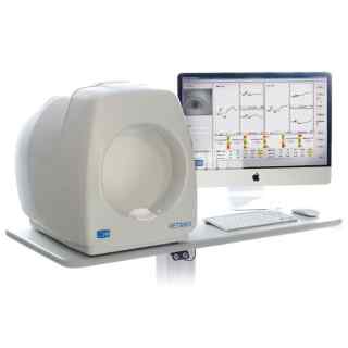
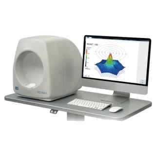
Electrophysiology testing includes a set of tests which can be used to provide information about the visual system beyond the standard clinical examination of the eye. Electroretinography (ERG) and Electro-oculography (EOG) are two of the tests that can be conducted by using the Retimax ocular electrophysiology systems from CSO.
The primary objective of the electrophysiologic examination is to assess the function of the visual pathway from the photoreceptors of the retina to the visual cortex of the brain. Information obtained from these diagnostic tests helps establish the correct diagnosis or may rule out related ophthalmic diseases.
Electrophysiologic testing is useful in diagnosing a variety of inherited retinal and optic nerve diseases, toxic drug exposure, inflammatory conditions, intraocular foreign bodies, and retinal vascular occlusions.
Full Field Electroretinography (ERG) is a test used to detect abnormal function in the retina (the light-detecting portion of the eye). Specifically, this test examines the function of the light-sensitive cells of the eye (the photoreceptors), and several other cells that process these light signals before they are sent to the brain. Photoreceptors are specialized nerve cells in the eye that are sensitive to light. There are two types of photoreceptors in the human eye: rods and cones. Cones provide central high-acuity vision used for reading and are also colour vision. There are 6 to 7 million cones in the retina, of which about 650,000 are concentrated in the foveola for central vision. Rods provide vision in dim light. There are about 120 million rods in the retina, of which none are located in the foveola.
During the ERG test, the photoreceptors produce tiny amounts of electricity in response to brief flashes of light. If ophthalmologists know exactly how much light enters the eye, and how much electrical response is generated, they could figure out how well the rods and cones are working. The differences in responses are analysed to differentiate diseases which affect the rods from those which affect the cones or other cells in the retina.
To detect the electrical response of both the rods and the cones, special electrodes are placed on the surface of the eye. There are several types of electrodes; the type of electrode used will depend on the type of test being performed, the age of the patient, and the suspected condition.


