Topographers
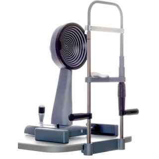
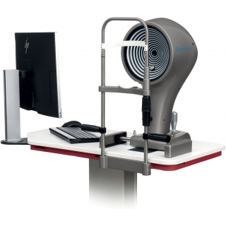
A topographer is an optical instrument designed to map the surface curvature of the cornea, non-invasively. Topography is also known as corneal topography, photokeratoscopy or videokeratography. The 3D map created by a topographer aids an ophthalmologist or optometrist in the diagnosis and treatment of numerous eye conditions and scenarios such as: pre cataract surgery, pre IOL implantation, pre LASIK surgery and contact lens fitting.
Corneal topography is exceptionably useful for examining characteristics of the cornea such as shape, curvature, power and thickness. It is also an essential tool for the contact lens specialist.
Over the past 150 years, the use of corneal topography has expanded from mapping of the cornea’s curvature to evaluation of several specific corneal and ocular surface characteristics. Practitioners can use it to assess the ocular surface prior to contact lens fitting, observe how a contact lens alters the shape and quality of the cornea and tear film, and monitor the relationship between the eye and the contact lens during wear.
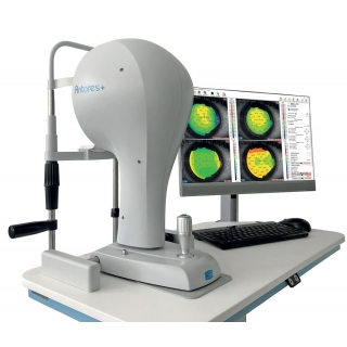
Among the most novel options on modern day topography instruments, like the new Antares Topographer from CSO, is the tear break-up displays.
Non-invasive tear break-up scores can be measured prior to initiating contact lens wear to gauge the quality of the natural tear film and see how it is affected by contact lens wear. A measurement is taken prior to lens wear and then compared to the subsequent measures to see how tear film quality is objectively affected in lens wearers with and without the lens in place.
Tear break-up displays can also be used when taking topography over the top of a contact lens. The surface wettability of the lens can be indirectly evaluated using this display, and a quantitative score will allow monitoring over wear time.
In addition, some instruments allow for video recording broken down by frames per second. This allows practitioners to dynamically evaluate changes in the tear film quality as patients blink.
The new Antares Topographer from CSO gives practitioners a full detailed dry report, based on the Ocular Surface Disease Index questionnaire (OSDI), limbal and conjunctival hyperaemia, Meibomian glands analysis, tear meniscus analysis, NIBUT, and tear osmolarity. This is then calculated, merging together all partial scores and provides an overall evaluation of the clinical condition of each patient and gives the practitioner a comprehensive diagnosis of dry eye.
As well as topographers being exceptionably useful for contact lens specialists and dry eye specialists, they are also useful for the following:
Keratoconus: Early screening of keratoconus suspects is one of the most useful roles of topography. Early keratoconus and suspects look normal on slit lamp examination, and the central keratometry (3 mm) gives only a limited assessment. Therefore, topography has become the gold standard in screening keratoconus suspects. In cases with established keratoconus, the role of topography is paramount for monitoring progression and doing a timely collagen cross linking, and in contact lens fitting.
Refractive surgery: To screen candidates for normal corneal shape, patterns and ruling out suspicious or keratoconic patterns. Post operatively, topography can help to assess the dioptric change created at corneal level (thus the effective change in the cornea), ruling out decentred or incomplete ablation, post excimer ectasia or other changes.
Post-surgery astigmatism: Post cataract surgery and post keratoplasty corneal astigmatism can be studied with the topographer and selective suture removal or other interventions can be planned.
Surgical planning in cases with astigmatism: Limbal relaxing incisions and other methods of topography guided incision placement are used by surgeons to reduce post-operative astigmatism.


